 Speaking of the unknown, we often look up to the sky, as if trying to see with our own eyes distant planets, star systems and galaxies. However, there is no need to travel thousands of light-years long when the unknown lies beneath our noses. Of course, we are talking about the oceans. Occupying a large part of our planet, the oceans are home to a wide variety of creatures: some live near the coast, others prefer hopeless darkness closer to the bottom. We do not know much about the inhabitants of the near-bottom layers of the oceans, but even less about the inhabitants of bottom rocks. Scientists from the University of Tokyo (Japan) have discovered a new species of bacteria that lives in numerous colonies in the ocean floor for many millions of years. Scientists believe that the study of these organisms will help find life on Mars. Where were the bacteria foundwhat are their features and how exactly does this discovery relate to life on Mars? The answers to these questions await us in the report of scientists. Go.
Study basis
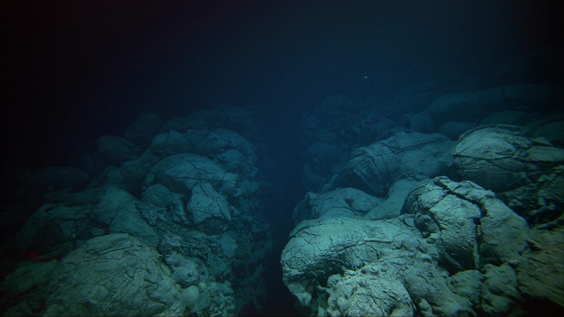
The upper layers of the oceanic crust consist mostly of basalts. The source of basalts is magma, which solidifies upon contact with air or water after an eruption. In the oceans, the main place of origin of basalts is the mid-ocean ridge - a string of ridges located in the central regions of all oceans. The height of these seamounts is about 2-3 km from the abyssal plains (plains of oceanic depressions). It is no secret that the process of basalt formation, being a high-temperature reaction, indirectly provides enough energy to maintain chemosynthesis, when inorganic compounds serve as fuel for the synthesis of organic compounds from CO 2 . This method of autotrophic (organic matter from inorganic) nutrition is unique to archaea (non-nuclear unicellular organisms) and bacteria.On the lateral sides of the ridge, the circulation of the crustal fluid is carried out hydrothermally within the basaltic lava, overloaded with sediments. The part of the basaltic lava under the sedimentary layer is called the basaltic base (foundation). Previous studies of ocean ridges 3.5–8 million years old showed that these relatively young aquifers * of the earth's crust are rich in anaerobic thermophiles * and aerobic mesophiles * , which are involved in the circulation of hydrogen, carbon and sulfur.Aquifer * - sedimentary rock from one or more underground layers of rocks with varying degrees of water permeability.
Thermophiles * - organisms that live in places where the temperature is above 45 ° C.
* — , , 20 45 °C.
When rock cracks are filled with secondary minerals, the intensity of fluid circulation and the reaction of basalt formation decreases, which corresponds to an increase in the age of the crust. Most of the oxidation occurs in the first 10 million years after the formation of the crust, and this is about 90% of the entire oceanic lithosphere of the Earth.According to scientists, given the vast area and inaccessibility of the ocean floor, a full study of its structure is an extremely complex process, even with current technologies. Nevertheless, there is evidence that the age of changes in the oceanic crust, indicating biological activity, is about 3.5 billion years. However, the role of microbial activity in the origin of these changes has not been evaluated previously. In this work, scientists describe the results of the analysis of samples obtained during the IODP expedition (integrated ocean drilling program from the integrated ocean drilling program), conducted in the South Pacific gyre (SPG from the South Pacific Gyre ).Analysis of the samples showed the presence of microbial cells in iron-rich smectite (a group of clay minerals that includes montmorillonite, nontronite, beidelite, etc.). Analysis of lipid profiles and DNA sequences of these organisms showed that they are heterotrophic bacteria, which indicates the presence of organic substances in underwater basalt.
Research results
In SPG, the deposition rate is quite low, from which sediments falling to the bottom are almost completely devoid of organic matter.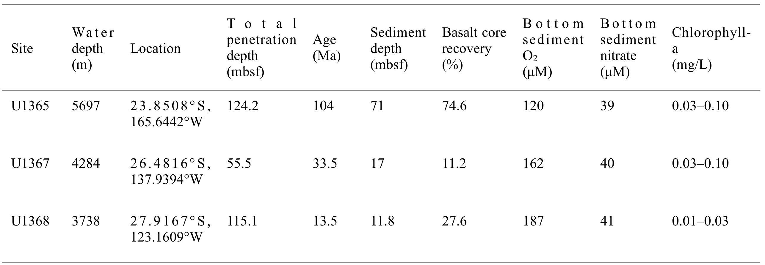 Table 1: sample data (mbsf - meters below the ocean floor; NS - no sampling; Ma - millions of years).Samples were taken in three regions with different ages: U1365 - 104 Ma; U1367 - 33.5 Ma; U1368 - 13.5 Ma.In such an ultra-oligotrophic environment *, dissolved O 2 penetrates from the ocean floor into the basalt foundation and supports the life of aerobic microbes throughout the entire depth of the deposits.
Ultra-oligotrophic environment * is an environment with an extremely low nutrient content.
First of all, the characterization of minerals from core samples with cracks was carried out to determine the presence of clay minerals, usually produced by low-temperature rock-water interactions (i.e. weathering). X-ray diffraction analysis revealed the presence of iron-rich smectite in core samples aged 33.5 Ma and 104 Ma, but not in core samples 13.5 Ma.Further, thin sections (U1367F-6R1, U1365E-8R4, and U1365E-12R2) corresponding to depths of 51, 109.6, and 121.8 mbsf (meters below the ocean floor) were prepared from these two samples. Sections were studied by scanning electron microscope (SEM) and transmission electron microscope (TEM). An energy dispersive X-ray spectroscopy (EDS) method was also employed .Iron-rich smectite was found in cracks filled mainly with celadonite oxyhydroxides in sample U1365E-8R4 and iron in U1365E-12R2. Whereas in sample U1367F-6R115, the cracks were filled with smectite rich in iron.In U1367F-6R1, two types of compositionally different Fe-rich smectite cracks were discovered:- the first type is similar to those found in U1365E-8R4 and U1365E-12R2 with a high content of Mg and K;
- the second type is characterized by a high iron content, which is usually observed in Fe-rich smectite from deep-water hydrothermal embankments.
Fluorescence microscopy of thin sections showed the presence of cellular fluorescence signals in the areas of cracks and veins associated with Fe-rich smectite in samples U1365E-8R4 and U1365E-12R2 (images below).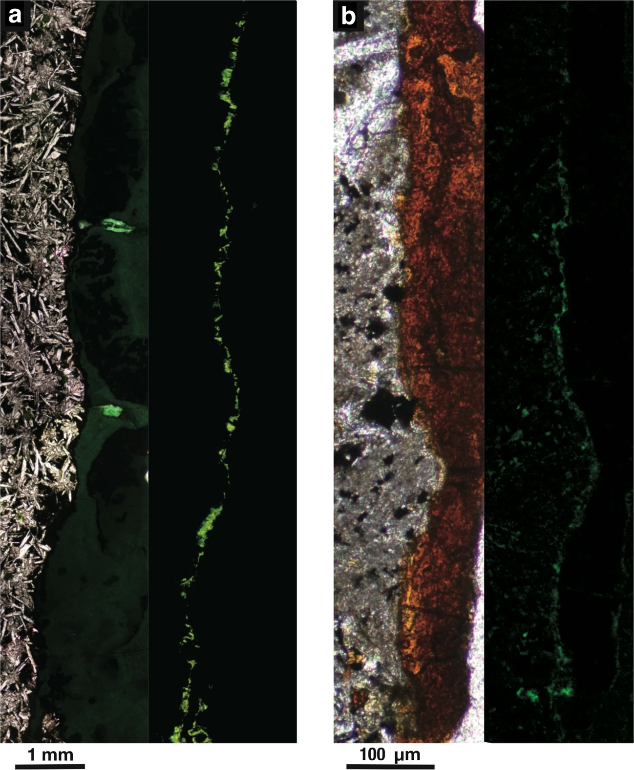 Image No. 1: images (light and fluorescence microscopy) of cells stained with SYBR Green I, in a crack filled with celadonite in U1365E-8R4 ( a ) and in a vein filled with iron oxyhydroxides in U1365E-12R2 ( b ).Although iron-rich smectite with a high Mg and K content in U1367F-6R1 corresponds to fluorescence signals, no fluorescence signals were detected in veins filled with iron-rich smectite in U1367F-6R1.To confirm that these greenish signals in the images come from microbial cells, and not from autofluorescent materials, 10 x 10 μm sections with a thickness of 3 μm were prepared using a focused ion beam for nano-sized secondary ion mass spectrometry (nanoSIMS).Analysis of U1365E-8R4 revealed overlapping 12C14N-, 31P-, and 32S- signals on dense spots stained with SYBR Green I, indicating that these greenish signals were obtained from microbial cells.
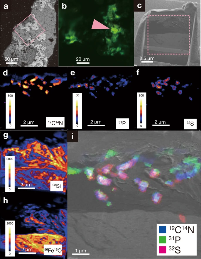
Image No. 2: Assessment of the cell population in cracked specimens:— U1365E-8R4;
b — , SYBR Green I;
— U1365-8R4 (1010 3 ), nanoSIMS;
d — NanoSIMS 12C14N-;
e — NanoSIMS 31P-;
f — NanoSIMS 32S-;
g — NanoSIMS 28Si-;
h — NanoSIMS 56Fe16O-;
i — - Ga ion image 1010 NanoSIMS 12C14N- , 31P- 32S- .
Microbial cells are localized in close proximity to microscale voids and are surrounded by iron-rich smectite. The same result was obtained for sample U1365E-12R2.The use of a transmission scanning electron microscope (PEM) confirmed that microbial cells are spatially connected with layers of iron-rich smectite. Given this connection and the large compositional difference between iron-rich smectite and bentonite clay used for drilling fluid, scientists concluded that microbial cells were not introduced from the drilling fluid, i.e. did not fall into the samples from the outside.The combination of these data suggests that numerous colonies of microorganisms live in the depths of the ocean floor (sorry for the pun). The next stage of the study was the determination of the microbial composition of these colonies.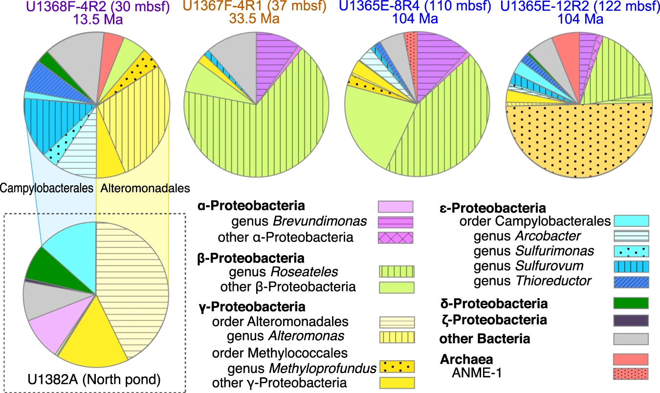 Image No. 3In accordance with the phylogenetic affiliation based on 16S rRNA gene sequences (Image No. 3), three types of microbial communities were identified:
- SPG-I ( : 13.5 ). U1368 γ- ε- , Arcobacter, Thioreductor, Sulfurimonas Sulfurovum (- / - *) Alteromonas ( *).
* — , .
* — , .
* — , .
- SPG-II (33.5–104 ). U1365 U1367 β-, *, Roseateles depolymerans.
* — , .
- SPG-III U1365E-12R2 ( 122 mbsf), γ-, Methylococaceae. Methylococaceae, , .
Microbial communities were previously observed in core and water samples from the North Atlantic region of the IODP expedition (core age 8 Ma), where oxygenated cold liquid actively circulates in the basalt foundation covered with sediments. However, there are clear differences in species. Microbial communities in the North Atlantic fluid samples are mainly composed of Campylobacterales and Alteromonadales . And in the new samples there was a significant advantage in the direction of Alteromonadales , while the representatives of Campylobacterales were in a clear minority.If we compare a sample from a relatively young site (8 and 13.5 Ma), then its microbial communities are almost identical to those found in samples from the North Atlantic site. Consequently, in the Atlantic Ocean and in the Pacific Ocean, the microbial communities found at the same depth of the bottom are almost identical.Knowing who lives in the bottom of the ocean, it was necessary to establish how these organisms survive there, that is, how they interact with the environment.The foundation at 13.5 and 33.5 Ma mainly consists of a lava cushion covered with deposits from 12 to 17 m thick. The deepest sediment in both sections contains the same concentrations of dissolved O2 and dissolved nitrate. Although the structure of the crust and the chemical composition of the dissolved oxidizing agent are quite similar in both areas, the composition of microbial communities noticeably differs in the 13.5 and 33.5 Ma foundations. Clay minerals that form in cracks / veins during low-temperature rock-water interactions provide information that can explain the difference between these communities.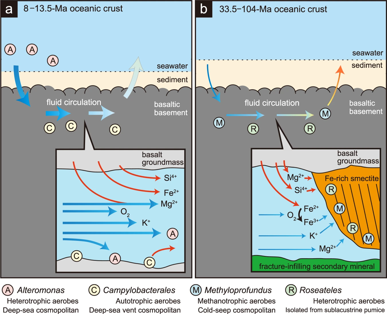 Image No. 4Thus, the presence and absence of iron-rich smectite in cracks / veins in sections U1367 and U1368 indicate that the formation of iron-rich smectite was inhibited by the active circulation of sea water in U136816.Although the basalt foundation at U1365 includes lava flows, where the fluid flow is usually between the sheet layers and not along the cooled edges of the lava pad, the composition of its microbial community is similar to that found at U1367, which is consistent with the presence of iron-rich smectite in U1365 and U1367.The heat flux at the bottom of U1367 and U1365 indicates thermal conductivity as the main method of heat transfer. While the heat flux on U1368 is not so active, it follows that here the main method of heat transfer is the circulation of fluid in the rocky crust.This difference is consistent with the age of the foundation in the respective areas, since fluid circulation and heat transfer are generally much more active in relatively young warm crust (such as the 13.5 Ma crust in U1368) than in the older and therefore colder bark (for example, bark 33.5 Ma in the area U1367 and 104 Ma in the area U1365).Scientists believe that the habitability of the foundation is controlled by heat and fluid flows, which tend to decrease over time compared to the primary structure of the crust. In addition, the formation of iron-rich smectite in the basaltic foundation appears to correlate with the species of microorganisms in the older oceanic crust.A more detailed examination of the 10x10 micron slice, which was mentioned earlier, showed that the cell density range isn x 3.3 x 10 9 cells / cm 3 , where n is the number of cells found in the slice.Therefore, in the samples U1365E-8R4 and U1365E-12R2, the approximate number of cells was 5.0 × 10 10 and 0.7 × 10 10 cells / cm 3 . Cell accumulation was limited to iron-rich smectite at the interface between basalt and other minerals.Within this interface, the cell density is extremely high compared to the cell density in the deepest sediment (~ 10 2 cells / cm 3) lying above the basalt foundations in sections U1365 and U136714, and compared with low-temperature liquids collected from a basalt foundation (8 Ma) from the North Atlantic region (~ 10 4 cells / cm 3 ).
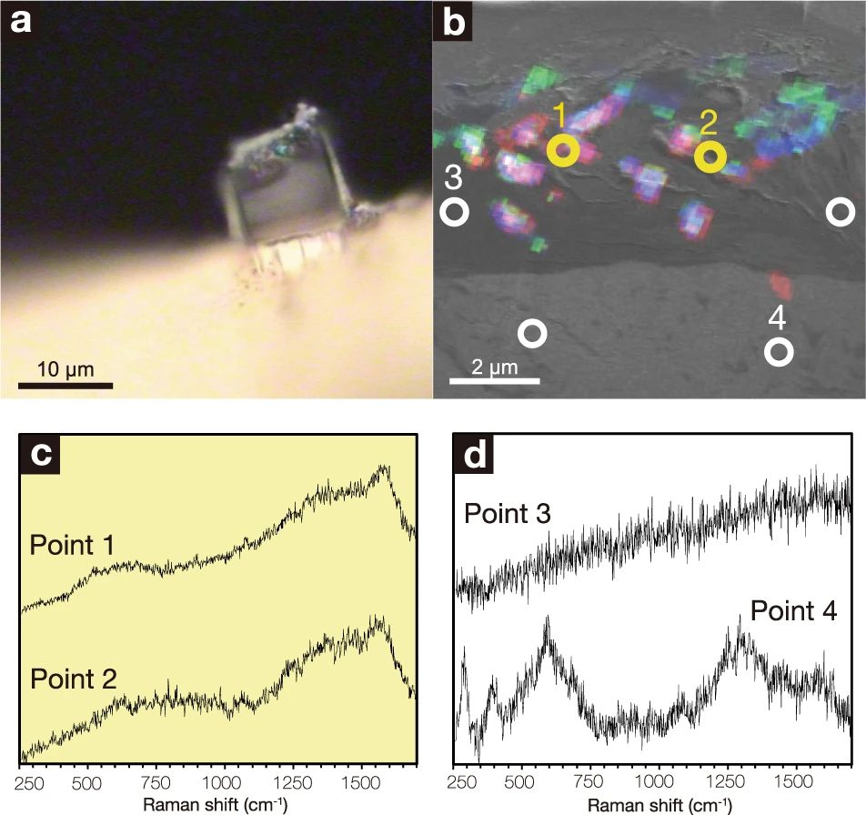 Image No. 5To verify the correctness of a certain cell density, µ-Raman spectroscopy was performed, which allows one to obtain a spectrum from microbial-smectite clusters (image No. 5).The spectrum was obtained at all interfaces filled with iron-rich smectite with a high Mg and K content in U1367F-6R1, U1365E-8R4 and U1365E-12R2, but not from U1367F-6R1 with iron-rich smectite and high Fe. The absence of a spectrum in iron-rich smectite may be due to its formation in a deep-water hydrothermal mound near the mid-ocean ridge.Smectite is a fine-grained clay mineral with a large surface area for the adsorption of organic substances. Since the dominant microbial communities found in basaltic bases 33 and 104 Ma are heterotrophic, it is possible that organic matter associated with iron-rich smectite helps maintain high cell density at the basaltic interface.Smectite samples taken for analysis contained 22 times more organic carbon than was found in other core sections. And this fully confirms the theory that organic matter associated with minerals stimulates the heterotrophic activity of microorganisms at the basalt interface.For a more detailed familiarization with the nuances of the study, I recommend that you look into the report of scientists and additional materials to it.
Epilogue
In this work, scientists studied ocean floor samples of different drilling depths, and therefore of different ages (104, 33.5 and 15.1 million years). The samples turned out to be rich in microorganisms, the density of which in some areas was about 0.7 × 10 10 cells / cm 3 . The ability to live in such unattractive conditions for bacteria is due to the clay mineral surrounding them - smectite.The rock contains cracks filled with smectite, which in turn concentrates the nutrients needed by bacteria.The results of this study not only help reveal some of the secrets of the ocean floor, but also contribute to the creation of new techniques for revealing life on other planets. The researchers themselves say that their find can be useful for such searches on Mars, since the mineral composition of the bottom of the oceans is likely similar to the mineral composition of the surface of Mars.Researchers intend to continue their work, but already in a team with NASA representatives. They plan to test their methodology on rock samples obtained from Mars.Thank you for your attention, remain curious and have a good working week, guys. :)A bit of advertising :)
Thank you for staying with us. Do you like our articles? Want to see more interesting materials? Support us by placing an order or recommending to your friends, cloud VPS for developers from $ 4.99 , a unique analog of entry-level servers that was invented by us for you: The whole truth about VPS (KVM) E5-2697 v3 (6 Cores) 10GB DDR4 480GB SSD 1Gbps from $ 19 or how to divide the server? (options are available with RAID1 and RAID10, up to 24 cores and up to 40GB DDR4).Dell R730xd 2 times cheaper at the Equinix Tier IV data center in Amsterdam? Only we have 2 x Intel TetraDeca-Core Xeon 2x E5-2697v3 2.6GHz 14C 64GB DDR4 4x960GB SSD 1Gbps 100 TV from $ 199 in the Netherlands!Dell R420 - 2x E5-2430 2.2Ghz 6C 128GB DDR3 2x960GB SSD 1Gbps 100TB - from $ 99! Read about How to Build Infrastructure Bldg. class c using Dell R730xd E5-2650 v4 servers costing 9,000 euros for a penny?